In celebration of 50 years of images captured by the light microscope, Nikon’s Small World competition is considered the premier platform for highlighting the beauty and intricacy of life as observed through a light microscope.
In celebration of 50 years of images captured by the light microscope, Nikon’s Small World competition is considered the premier platform for highlighting the beauty and intricacy of life as observed through a light microscope.
The Photomicrography Competition invites anyone passionate about microscopy and photography to participate. Additionally, the Small World In Motion video competition includes any films or digital time-lapse photography captured through the microscope.
The Nikon Small World Competition was launched in 1975 to honor and celebrate the contributions of those engaged in photography through the light microscope. Since its inception, Small World has evolved into a premier platform for photomicrographers across a diverse range of scientific fields.
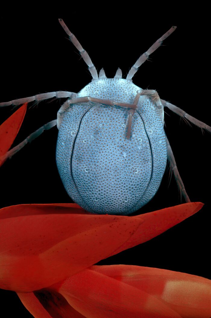
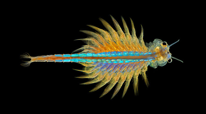
1
A Water Mite - Jacek Myslowski, Wloclawek, Kujawko-Pomorskie, Poland
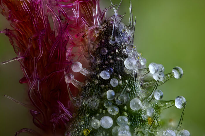
2
Part of a Cannabis plant's Bract, the plant's reproductive structures and the bulbous glands known as are trichomes - Chris Romaine, Port Townsend, Washington, USA
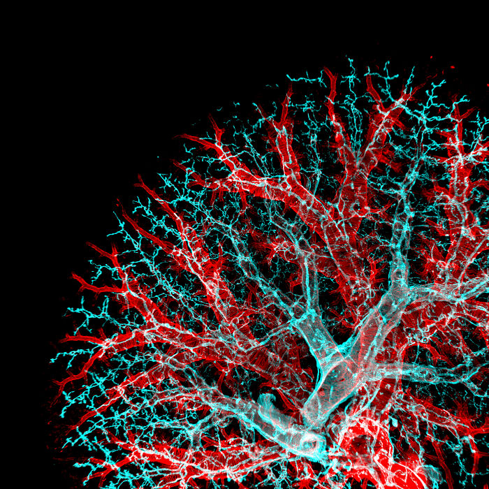
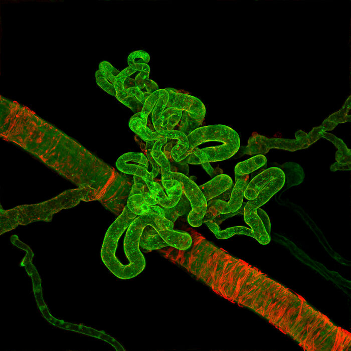
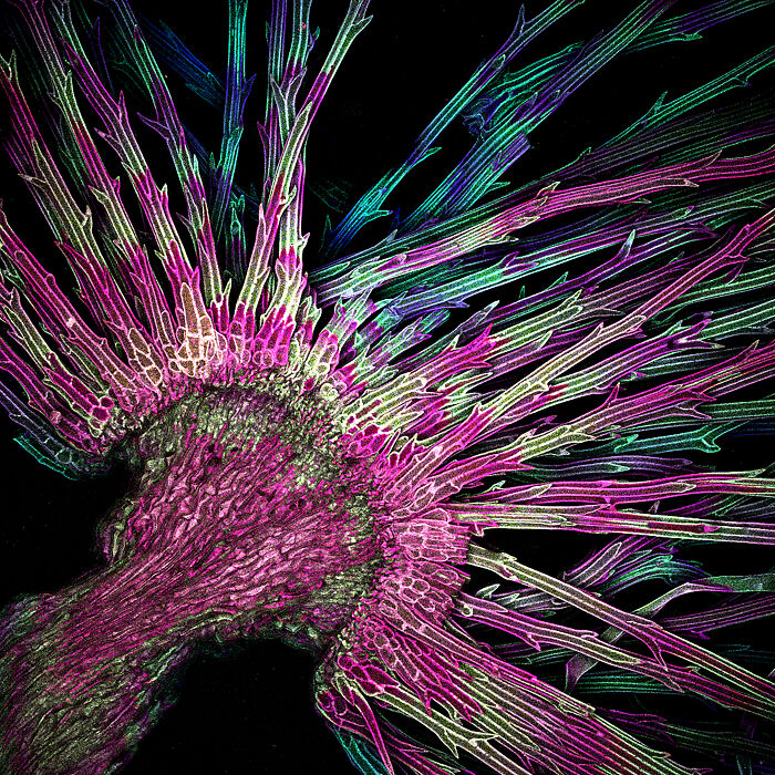
3
Some Lymphatic vasculature (cyan) and vessels (red) of a mouse lung - University of California, San Francisco Pulmonary, Critical Care, Allergy and Sleep Medicine, San Francisco, California, USA
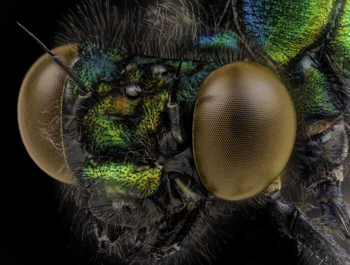
4
A Yong male Damselfly - Oregon Department of Agriculture (ODA) Entomology Lab Albany, Oregon, USA
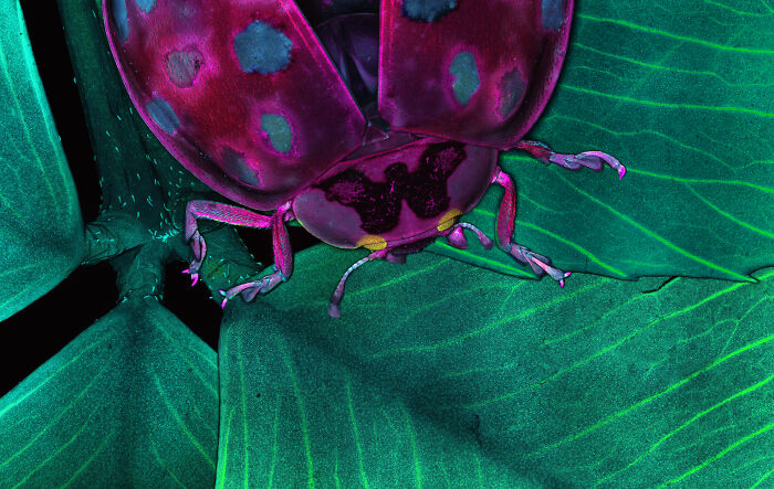
5
A Ladybug on a clover leaft - MDI Biological Laboratory Murawala Lab, Bar Harbor, Maine, USA
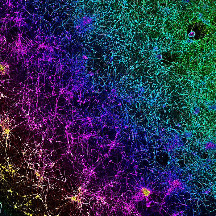
6
A network of dopaminergic neurons generated from human stem cells - University of Oxford Nuffield Department of Clinical Neurosciences (NDCN) Oxford, Oxfordshire, United Kingdom
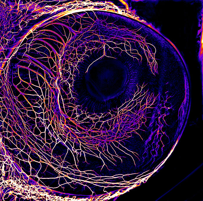
7
The Developing nervous system in the eye of a 7-day-old chick embryo - University of Zurich Department of Molecular Life Sciences Zurich, Switzerland
8
An Abnormal blood vessel formation in a human retina with severe diabetic retinopathy - Lions Eye Institute Physiology and Pharmacology laboratory, Nedlands, Western Australia, Australia
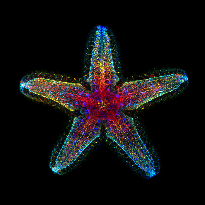
9
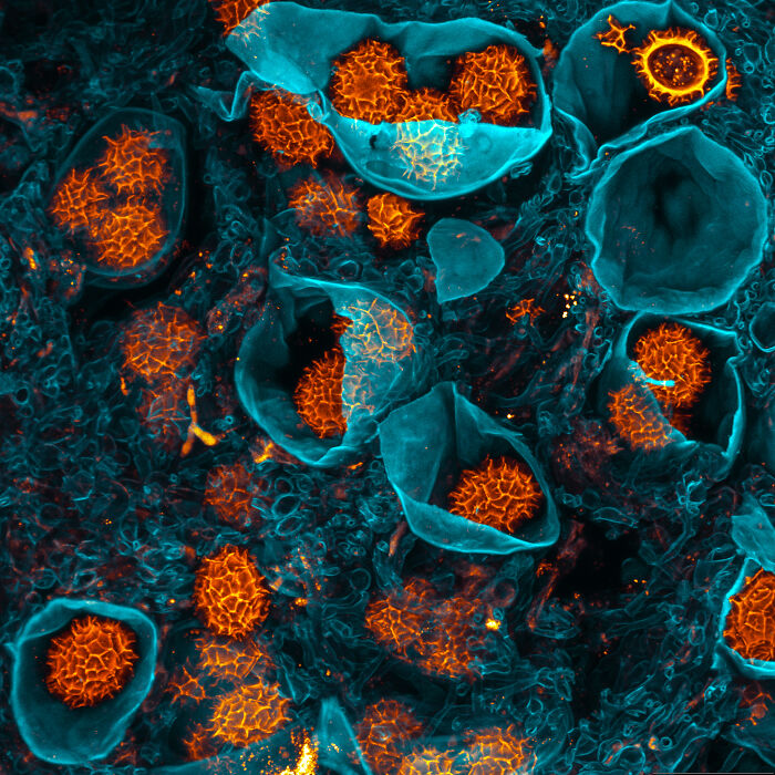
The Nervous system of a young Star Fish - Stanford University Department of Molecular and Cell Biology Pacific Grove, California, USA
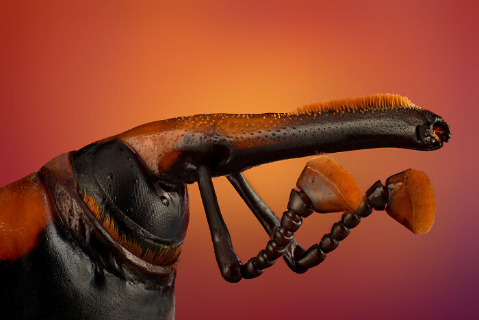
10
The anterior section of Palm Weevil - Tanta University, Faculty of Science Department of Zoology Tanta, Egypt, Arab Republic
11
A Dandelion pappus - Amicus Therapeutics, Philadelphia, Pennsylvania, USA
12
A Moss sporophyte with green spores - Joshua Coogler Dallas, North Carolina, USA
13
The Cross section of a leaf of European beach - Maria Enzersdorf, Austria
14
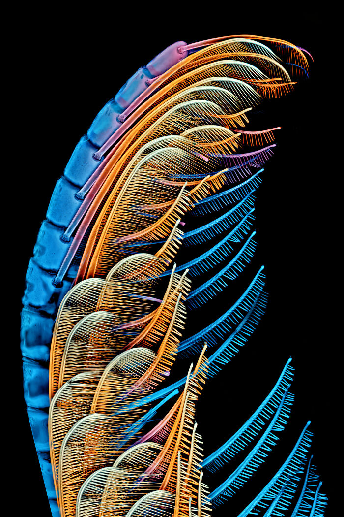
The Antenna of a mole crab - Howard Hughes Medical Institute (HHMI), Janelia Research Campus Ashburn, Virginia, USA
15
A section of small intestine of a Mouse - Medical University of South Carolina Department of Regenerative Medicine & Cell Biology, Charleston, South Carolina, USA
16
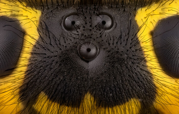
The Ocelli between the compound eyes of a Yellow Jacket - Dr. Bruce Douglas Taubert, Glendale, Arizona, USA
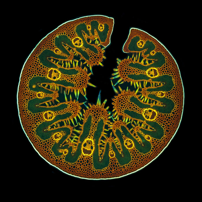
17
The cross section of a Dandelion showing curved stigma with pollen - University of Nottingham School of Life Sciences, Super Resolution Microscopy Nottingham, Nottinghamshire, United Kingdom
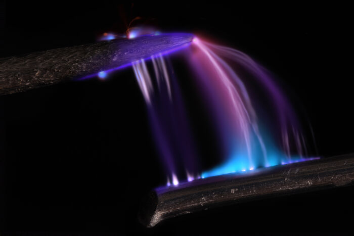
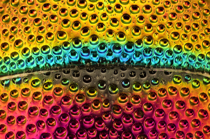
18
The Electrical arc between a pin and a wire - Dr. Marcel Clemens, Verona, Veneto, Italy
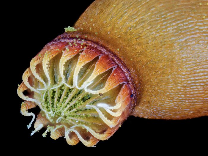
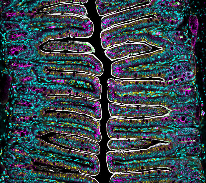
19
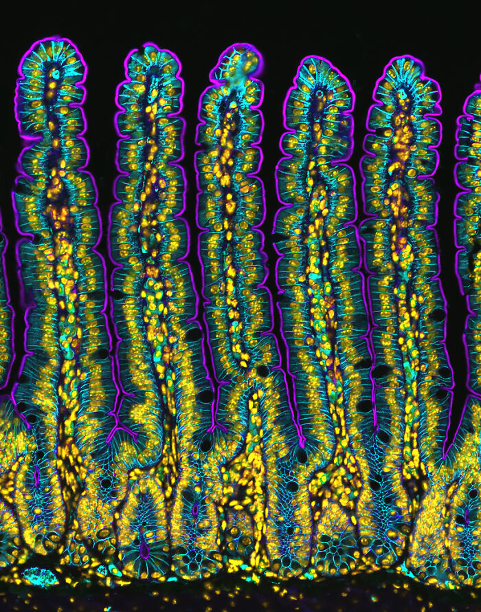
Intestinal villi - Medical University of South Carolina Department of Regenerative Medicine & Cell Biology, Charleston, South Carolina, USA
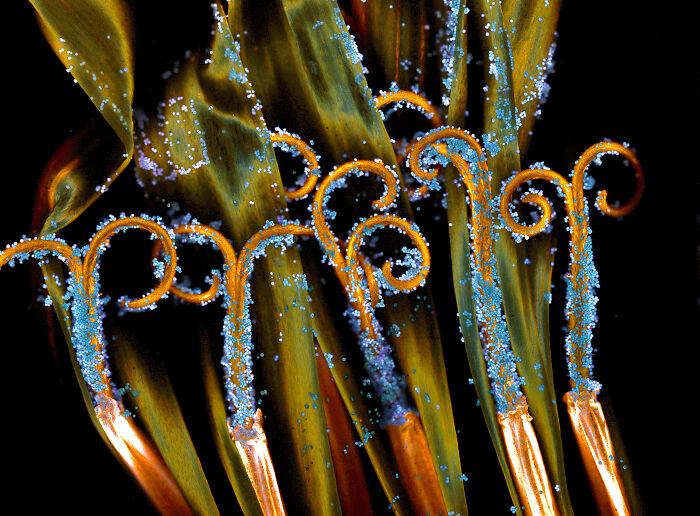
20
A Cross section of a beach grass leaf - Gerd Gunther, Düsseldorf, Germany
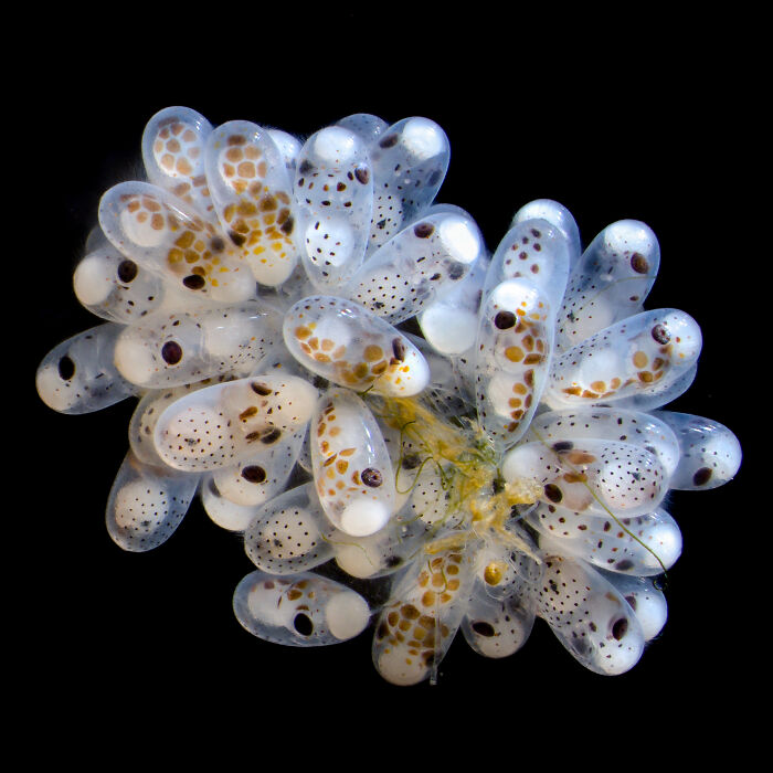
21
A Cluster of Octopus eggs - Columbia University Department of Neurobiology and Behavior New York, New York, USA
22
The Dorsal part of Cuckoo Wasp's abdomen -Daniel Knop, Oberzent-Airlenbach, Hessen, Germany
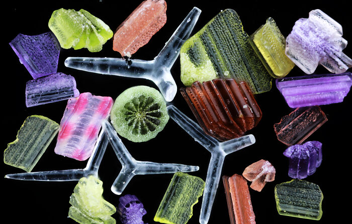
23
Beach Sand - National Astronomical Observatories, Chinese Academy of Sciences Beijing, China
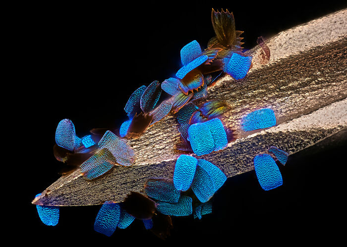
24
The scales on the Wing of a butterfly on a medical syringe - Oberzent-Airlenbach, Hessen, Germany
25
A Brine Shrimp - Christopher Algar, Hounslow, Middlesex, United Kingdom